Disclaimer: Due to unforeseen difficulties, we have had to take down the images on this notes page. They will be replaced shortly. We apologise for the inconvenience, but hope that the new images will provide you with an even better learning experience.
- Describe the circulatory system as a system of tubes with a pump and valves to ensure one-way flow of blood.
It’s a pretty self-explanatory point. The tubes are our blood vessels (veins, arteries, etc.), the pump is the heart, and we have valves in our veins and heart to ensure that blood won’t flow backwards.
- Describe double circulation in terms of a low-pressure circulation to the lungs and a high-pressure circulation to the body.
Double circulation means that the blood flows through two circuits – one low-pressure one and one high-pressure one.
The low-pressure circuit is when the blood travels from the heart to the lungs, and back. This is also known as the pulmonary circuit.
The high-pressure circuit, or systemic circuit, is when blood flows from the heart to the rest of the body, and back. This is higher pressure because the blood has to travel further, so the heart applies a greater force on this blood.
- Describe the structure of the heart, including the muscular wall and septum, atria, ventricles, valves and associated blood vessels.
So, the heart actually looks like this:
But, here’s a simplified diagram to make it easier to understand:
Before we launch into the explanation, you should note that all the diagrams here are not a mirror reflection of your own heart – they are drawn so that it is like looking at someone else’s heart.
I.e. the right side of the heart on the diagram, will be on your left.
The heart is separated into two, by a large muscular wall down it’s centre called the septum.
Both of these sides are further divided into two chambers, the top two chambers being the atria (sing. Atrium) and the bottom two being the ventricles. This gives us the right atrium, the left atrium, the right ventricle and the left ventricle.
The atria and ventricles are separated by the atrio-ventricular valves. These open and close to control the direction of blood flow.
The valve on the right side is the tricuspid valve (because it has three cusps, or flap-like things) and the valve on the left is the bicuspid valve.
The venacava is a vein that brings blood into the right atrium; the superior venacava is the branch of the venacava that comes from the top and the inferior venacava is the branch that comes from the bottom.
The pulmonary artery takes blood from the right ventricle to the lungs, and the pulmonary vein takes blood from the lungs to the left atrium. The aorta is an artery that takes blood from the left ventricle to the rest of the body. Note that these arteries have their own valves too, to prevent the backflow of blood. The one in the pulmonary artery is the semi-lunar pulmonary valve and the one in the aorta is the aortic semi-lunar valve.
- Describe coronary heart disease in terms of the blockage of coronary arteries, and state the possible causes (diet, stress, smoking and genetic factors) and preventive measures.
Coronary heart disease (CHD) is caused by artherosclerosis.
This is when plaque builds up in your arteries, thereby narrowing or blocking them up. This plaque is made of cholesterol, fatty substances, cellular waste products, calcium and fibrin.
This buildup of plaque usually takes several years.
Overtime, the plaque may harden, reducing the flow of oxygen rich blood to the heart muscles. Sometimes, this plaque might rupture (break apart), causing the formation of blood clots, or the broken piece of plaque to travel down to a narrow arteriole and block it up. Both of these completely cut off the supply of oxygen to the heart muscles. This could cause heart failure.
This, is coronary heart disease.
CHD can be caused by a diet high in fat, especially saturated fats, it can be caused by stress, smoking, and sometimes, it is hereditary.
In order to avoid CHD, it is important to maintain a balanced diet, have plenty of exercise, avoid unhealthy habits like smoking. Taking time to yourself and relaxing also helps to reduce stress, and thereby, reduce the risk of CHD.
- Describe the function of the heart in terms of muscular contraction and the working of the valves.
When the entire heart is relaxed, the heart is in diastole. During diastole, the pulmonary and aortic semilunar valves, and the atrioventricular valves are open. During diastole, the atria are filled with blood.
Next is atrial systole:
In this stage, the muscular walls of the atria contract, squeezing blood into the ventricles. The atrioventricular valves remain open, but the semilunar valves are pushed shut due to the pressure of the blood.
Finally, in ventricular systole, the relax and the ventricular walls contract. This causes the atrioventricular valves to close and the semilunar valves to open, so blood is pushed out into the aorta and pulmonary artery.
Then, ventricular diastole occurs again, and so the cycle restarts.
- Investigate the effect of physical activity on pulse rate.
Measure the pulse rate of a person at rest.
This can be done by pressing two fingers down on the inside of their wrist, between the bone and the tendon on their thumb side. You should be able to feel pulses. This is because, during ventricular systole, blood is forced down the arteries, so they expand slightly. Using a stopwatch, count the number of pulses in 30 seconds, and multiply by two to gain the pulse rate per minute.
That person should then undergo exercise, e.g. jogging for 1 – 5 minutes.
Measure their pulse rate again, and compare.
- Investigate, state and explain the effect of physical activity on pulse rate.
Physical activity means the increased usage of muscles. As the muscles are working more, they require more energy, and hence, need more oxygen. This means that more blood needs to be pumped to the muscular tissue, so the heart works faster, hence increasing the pulse rate.
(pulse rate is a measure of the number of heart beats per unit time, it’s usually measured as the number of heart beats per minute.)
- Name the main blood vessels to and from the heart, lungs, liver and kidney.
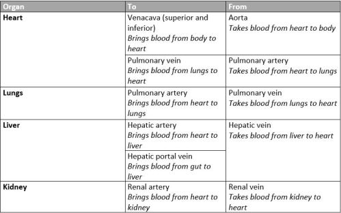
- Describe the structure and functions of arteries, veins and capillaries.
Arteries have the thickest walls. The elastin in their walls allow them to expand to accommodate the blood during each pump of the ventricle (instead of bursting), and it allows them to recoil to push the blood and maintain the high pressure.
Veins have much thinner walls, so their tunica media and tunica externa are relatively thin. As opposed to arteries, veins have valves to prevent the back flow of blood, especially when blood has to flow against gravity. The muscles around veins contract and relax, providing a force to push blood along the veins.
Capillaries are the smallest blood vessel, and carries the lowest pressure blood. Usually, only one RBC can fit through the diameter of the capillary (Capillaries usually have a diameter of approximately 7 micrometres, and RBCs have a diameter that is approximately equal to that.). This means they only have one layer, instead of three.
All three blood vessels have a single layer of endothelial cells (squamous epithelium). Arteries and veins have a tunica media (smooth muscle) and a tunica externa (elastin and collagen). Arteries have more elastin than veins in their tunica externa.
Arteries have a relatively small lumen (space inside the vessel), and veins have a larger lumen. Capillaries have the smallest lumen.
- Explain how the structure and function are related to arteries, veins and capillaries.
Arteries have the highest-pressure blood flowing through them, so they have the thickest walls. They don’t need valves.
Veins have blood at a lower pressure flowing through them, so their walls don’t have to be as thick. They also have a much larger lumen, allowing blood to flow much more easily through them. Veins need valves to prevent the back flow of blood.
Capillaries are very small in diameter, allowing them to bring blood closer to the needy tissues. This reduces the diffusion distance of oxygen from RBCs to tissue, and carbon dioxide from the tissue to the RBCs. They have a single cell thick wall, again reducing diffusion distance, and blood flows relatively slowly through arteries, allowing more time for diffusion.
Capillaries form an extremely extensive network of blood vessels, so despite individual capillaries having a small cross sectional area, capillaries have the largest total cross sectional area of all three blood vessel types.
- Identify red and white blood cells as seen under the light microscope on prepared slides, and in diagrams and photomicrographs.
Red Blood Cells:
White Blood Cells:
White Blood Cells, also known as leukocytes, are a little bit more complicated, as there are many types of WBCs.
There are 5 main types of WBCs –
- Monocytes, which later mature into macrophages – They have a long lifespan and help kill bacteria.
- Lymphocytes – These create antibodies that play an important role in the battle against foreign bodies like bacteria and viruses.
- Neutrophils – These are the most common WBC (50 – 70% WBCs in circulation are neutrophils). They digest and kill bacteria by phagocytosis.
- Basophils – they ‘sound an alarm’ by secreting chemicals, to alert other WBCs of infectious pathogens.
- Eosinophils – They attack and kill parasites, destroy cancer cells, and help with allergic responses.
These WBCs are often sorted into two groups called granulocytes (the WBCs with granules in their cytoplasm) and agranulocytes (the WBCs without granules).
The granulocytes are neutrophils, eosinophils and basophils.
The agranulocytes are monocytes and lymphocytes.
They are also sorted into two different groups – lymphocytes (the cells that produce antibodies) and phagocytes (the cells that perform phagocytosis).
Neutrophils are characterised by multiple lobes on their nuclei, and granules in their cytoplasm.
Eosinophils have two lobes on their nuclei and granules.
Basophils have two lobes on their nuclei again, are usually stained purplish-black, and have granules
Lymphocytes are the smallest of the WBCs and have a large spherical nucleus that takes up most of the cytoplasm.
Monocytes have a kidney shaped nucleus and plenty of cytoplasm.
- List the components of blood as red blood cells, white blood cells, platelets and plasma.
Blood is made up of red blood cells, white blood cells, platelets and plasma,
- State the functions of blood:
- Red blood cells – haemoglobin and oxygen transport
Red Blood Cells are made of many thousands of haemoglobin, each of which are made of four polypeptides. Each polypeptide has one iron ion (Fe2+) attached to it. This is where an oxygen molecules binds. This means, each haemoglobin molecule can carry up to 4 oxygen molecules and hence, 8 oxygen atoms.
When the RBCs are in the lungs, they are surrounded by a high concentration of oxygen, leading to more and more oxygen binding with the haemoglobin. This leads to the blood becoming saturated with oxygen – it is carrying its maximum amount of oxygen. As the RBCs leave the lungs, and are transported in blood to respiring tissue, they are transporting oxygen to these tissues. As these tissues use up oxygen, the RBCs are surrounded by less oxygen, resulting in the release of oxygen from the haemoglobin. This is possible because the bond between the oxygen molecule and the haemoglobin isn’t strong enough to be permanent.
- White blood cells – phagocytosis and antibody formation
Neutrophils, macrophages, eosinophils and basophils can perform phagocytosis, and so, they are called phagocytes.
However, if they ask which WBC performs phagocytosis in a paper, the expected answer is usually neutrophils.
Phagocytosis is the ingestion and digestion of bacteria by white blood cells. This successfully breaks down the pathogen into its harmless components.
The stages of phagocytosis:
- ingestion: the bacteria/ food particles are engulfed by the WBC. This results in the formation of a food vacuole.
- Vesicles, called lysosomes, containing digestive enzymes, fuse with the food vacuole, dumping the enzymes into said vacuole.
- The bacteria are digested.
- The components of the bacteria are often egested (dumped outside the WBC).
Antibodies are formed by lymphocytes.
On the left is a diagram of an antibody. The Fv has different shapes in different antibodies – each type of Fv can bind with a different type of bacterium. This ensures that each different bacterium can be binded to by an antibody. The Fc is the same in every antibody.
The functions of antibodies include:
- Act as a label: Cells with antibodies binded to them can be identified as target cells by phagocytes.
- Neutralisation: the binding of an antibody to a pathogen can often cause the neutralisation of harmful toxins they release.
- Agglutination: They may cause several pathogens to stick together, preventing them from dividing and multiplying, causing them to die out.
- Platelets – causing clotting (no details)
Platelets are small disc shaped cell fragments, that are involved in blood clotting.
- Plasma – transport of blood cells, ions, soluble nutrients, hormones and carbon dioxide.
About 55% of blood is plasma. This is the solution that carries blood cells, and other solutes around the body.
Plasma is a pale-yellow sticky liquid. It is 92% water, 8% dissolved protein, soluble nutrients, hormones and carbon dioxide.
It takes RBCs close to respiring tissue to supply them with oxygen, it takes carbon dioxide from the respiring tissue and to the lungs, it carries nutrients from their sites of production to their sites of usage or storage, and transports hormones to their target organs.
- Describe the immune system in terms of antibody production, tissue rejection and phagocytosis.
The immune system is the body’s system of defence against potentially dangerous foreign bodies – kind of like its army.
Lymphocytes produce antibodies that label, neutralise and agglutinate pathogens.
Phagocytes digest and kill potentially dangerous pathogens.
Tissue rejection:
Tissue rejection is when a foreign tissue is placed in the body – e.g. during organ transplants – and is rejected by the body.
The body recognises these cells as foreign, and hence, attacks it by antibody production and phagocytosis. This is often a problem during tissue transplants, so patients are often given immunosuppressant tablets (tablets that suppress their immune system), once they undergo a transplant.
Notes submitted by Sarah.
Click here to go to the next topic.
Click here to go to the previous topic.
Click here to go back to the Science menu.
